11 February 2008

Nerve injury

การบาดเจ็บของเส้นประสาท(Nerver injury)ที่แบ่งโดย Seddon ในปี 1943 แบ่งได้เป็นสามแบบ

nerve fiber

  1. Neurapraxia คือ เป็นอาการบาดเจ็บที่รุนแรงน้อยที่สุด ส่วนใหญ่มักเกิดเส้นประสาทโดนกด ทำให้การนำสัญญาณของเส้นประสาทเสียไป เมื่อหายดีแล้วก็จะส่งสัญญาณประสาทได้ตามปกติ การบาดเจ็บแบบนี้ไม่ทำให้เกิด degeneration
  2. Axonotmesis คือ การขาดของ axon (nerve fiber) โดยที่ endoneurium ยังไม่ขาดออกจากกัน การบาดเจ็บแบบนี้จะทำให้สูญเสียความรู้สึก และไม่สามารถบังคับอวัยวะให้เคลื่อนไหวได้ แต่การบาดเจ็บแบบนี้ยังสามารถหายได้ เนื่องจาก endoneurium ยังอยู่ axon สามารถงอกใหม่ไปตาม endoneurium ได้
  3. Neurotmesis คือการขาดของเส้นประสาท โดยที่ endoneurium ก็ขาดไปด้วย การบาดเจ็บแบบนี้เป็นการบาดเจ็บที่รุนแรงที่สุด แก้ไขได้ด้วยการผ่าตัดเท่านั้น
Relate posted

05 February 2008

Localized teeth with radiopraph (x-ray)

One method to localized teeth(object) in dental radiograph is SLOB rule.

SLOB is shorten from Same Lingual Opposite Buccal.

Let take a look at the picture below to explain this rule.

same lingual opposite buccal x-ray

The object at lingual site will move in the same direction of the x-ray tube, opposing to the buccal object.

This rule also works in vertical dimension.

03 February 2008

Maxillary Fracture

  1. Fracture Zygoma (Tripod Fracture) น่าจะเป็น fracture ที่เกิดขึ้นมากที่สุดในบรรดา fracture of facial bone pattern ของ fracture มักจะเกิดจาก direct blow to body of zygoma จากการตรวจจะพบเห็นลักษณะของ contour abnormalities,มีอาการของ extraocular muscle entrapped (คือมี limitation of eye movement ในบางทิศทาง) และ force ที่มาจากทาง lateral wall of orbit อาจส่งผลไปถึง orbital apex ,optic canal เกิด diminish vision ได้ ,นอกจากนั้นการ displace ไปของ zygoma อาจไปกีดกัน motion of mandible

    ส่วนในภาพรังสี x-ray โดยเฉพาะใน water’s view จะเห็น discontinuity lines of Dolan และแน่นอนครับ ผู้เขียนแนะนำว่า CT จะช่วยการ diagnosis ได้ดีมาก


  2. Fracture Zygomatic arch pattern of fracture นั้นเกิดจากมีแรงมากระทำทางด้านข้างของ ใบหน้า ลักษณะของใบหน้านั้นจะพบว่ามี flatness of lateral cheek area and inability to open mouth อันเนื่องมาจากการที่ zygomatic arch fragment ลงมาขัดขวางการเคลื่อนที่ของ coronoid process และ temporalis muscle ส่วนภาพรังสีที่ใช้ในการ diagnosis ได้ดีคือ view submentovertex


  3. Fracture of Alveolar process of Maxilla เกิดขึ้นกับ small piece of maxilla ร่วมกับมี several fracture teeth สำหรับข้อ comment เกี่ยวกับ x-ray นั้นมีอยู่ว่าในกรณีที่สงสัย film chest x-ray จะช่วยในการตรวจสอบว่ามีการ aspirate foreign body หรือ tooth fragment ลงไปใน airway หรือไม่


  4. Orbital floor fracture , or “Blowout” fracture mechanism ของ fracture ก็คือว่า มีแรงหรือวัตถุมากระแทกรอบๆบริเวณดวงตา แล้วส่งแรงผ่าน soft tissue กระจายไปรอบๆดวงตาและผ่านมายัง thin floor of orbit เกิดการ fracture floor orbit และมี dropdown of orbital content to maxillary sinus

    clinical sign ที่พบได้ก็คือ enophthalmos and diaplopia ส่วนใน film Water’s view จะพบเงาของ soft tissue mass บริเวณ superior margin of maxillary sinus หรืออาจจะเห็น trapdoor fragment of bone protruding down to sinus


  5. Fracture Nose โดยปกติแล้วเราสามารถเห็น fracture นี้ได้จาก standard lateral skull film แต่ถ้าเราใช้ technique low kVp ใน x-ray ก็จะสามารถเห็น บริเวณ fracture นั้นและ soft tissue ได้ดีขึ้น แต่ก็ต้องระวังการดู film ผิดพลาดคือเห็น normal suture line หรือ linear channel for Nasociliary nerve เป็น fracture line ได้เหมือนกันนะครับ เราควรจะดูใน lateral film ที่มีแนวรังสีตั้งฉากกับ nose จะทำให้เห็น fracture line ที่มักจะเป็นแนวตั้งฉากกับ nasal bridge ได้ชัดเจนกว่า


  6. LeFort fracture หรือเป็น fracture ที่ complex bilateral fractures associated with large unstable fragment and invariably involve pterygiod plates โดยแนวของ fracture นั้นแบ่งเป็น 3 ระดับคือ

    • LeFort I มีแนว Fracture ตาม transmaxillary plane

    • LeFort II มีแนว fracture ตาม subzygomatic หรือ pyramidal plane ที่มี apex บริเวณ nasal bridge

    • LeFort III มีแนว fracture ตาม craniofacial plane


    6.1 LeFort I หรือ tranmaxillary fracture มีแนวหัก ไปตาม maxillary floor & orbital floor โดย involve medial & lateral walls of maxillary sinuses และทางด้านหลัง involve pterygoid plate of sphenoid bone จะพบลักษณะ floating maxilla

    6.2 Lefort II หรือ เรียกตามแนวทางของ fracture line ว่า Pyramidal fracture ลักษณะของ fracture นี้หลังจากมีแรงในแนวตรงเข้าด้านหน้ามากระทำแล้วจะมี mechanism ที่ downward below to nasal area

    6.3 LeFort III เป็น most severe ของ LeFort fracture ลักษณะจะเป็น Large unstable(floating) fragment ทั่วไปทั้งใบหน้า หรือเรียกตามอาการว่า Craniofacial disassociation

  7. Smash Fracture มีการคิดคำนี้ขึ้นมาเพื่อใช้เรียก severe comminution of the face with underlying skull injuries และยังรวมไปถึง fracture facial bone ที่ไม่สามารถ classified ไว้ในกลุ่มที่ผ่านๆมาได้ โดยรวมไปถึง Fracture frontal ,naso-ethmoidal ,naso-frontal ซึ่ง Fracture ในกลุ่มนี้ควรใช้ CT ในการตรวจจะได้รายละเอียดที่ดีกว่า plain film ทั่วไป
Article form http://www.geocities.com/omfsesan/facialfracture.htm

Relate links
Diagnosis Image Facial Fracture
Mandibular fracture

Mandibular fracture

Common site of Mandibular Fractures

mandibular fracture type

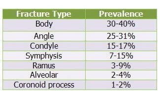
Mandible นั้นถือเป็น prominent position ที่มักเกิดการบาดเจ็บได้ง่าย ในสมัยก่อน นั้นมีอุบัติการณ์ประมาณ 2:1 เมื่อเทียบกับ facial bone fracture อื่นๆ แต่หลังจากมี high speed automobile accident มากขึ้น อุบัติการณ์ก็มาใกล้เคียงกัน โดย fracture mandible มี Clinical sign ทั่วๆไปหลายอย่างเช่น facial distortion, malocclusion, abnormal mobility, stepping formation เป็นต้น

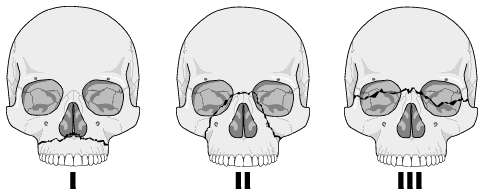
โดยปกติแล้วเราถือ fracture mandible อยู่ใน” Ring bone rule” คือกระดูกที่มีลักษณะโค้ง ดังนั้นเมื่อเรา เห็นแนว fracture แห่งหนึ่งจากการตรวจหรือจากภาพรังสีก็ตามทีแล้ว ก็มีโอกาสสูงที่เราจะพบ fracture อีกแห่งหนึ่งในmandible ด้านตรงกันข้าม เนื่องจากลักษณะการกระจายแรงที่มากระทำของกระดูก mandible นี้ นอกจากนั้นยังมีปัจจัยการมี flexibility ของ รูปร่าง mandible รวมไปถึง การ absorb แรงโดย TMJ

จากกฎข้างต้น Fracture mandible นั้นจึงอาจเกิดเป็น double fractures ได้เช่น bilateral sides of symphysis ,combination include angle mandible plus contralateral body or condyle ส่วน triple fracture นั้นก็อาจเกิดขึ้นได้เช่น fracture both condyle and symphysis ครับ

สุดท้ายมีข้อคิดแถมอยู่ใน website นะครับ - Wise saying about Mandibular fracture

  1. คำนึงถึง “Ring bone Rule” เอาไว้
  2. Panoramic film นั้นนับเป็น single film ที่ใช้ตรวจดู fracture mandible ได้ดีมาก
  3. สังเกตโดยรอบของ cortical margin of mandible เพื่อหา discontinuities( fracture line) line
  4. อย่าลืมสังเกตดูว่ามี discontinuities of inferior alveolar canal หรือไม่
  5. Fracture line ที่ผ่าน root นั้นเรานับเป็น open fracture
  6. Mandibular Fractures นั้นยังมีสาเหตุหลายอย่างที่มากกว่า trauma เช่น Pathologic fracture นั้นสามารถเกิดขึ้นได้ จาก ทั้ง tumor, infection เช่น osteomyelitis
Article from http://www.geocities.com/omfsesan/facialfracture.htm

Relate links
Diagnosis Image Facial Fracture
Maxillary fracture