This post will show the radiographic sign that I tell you in x-ray and fracture line posted
The figure below shows the oblique fracture that the fracture fragment was oblique and overlapped in bucco-lingual direction, thus, show the more radiopaque line (red arrow)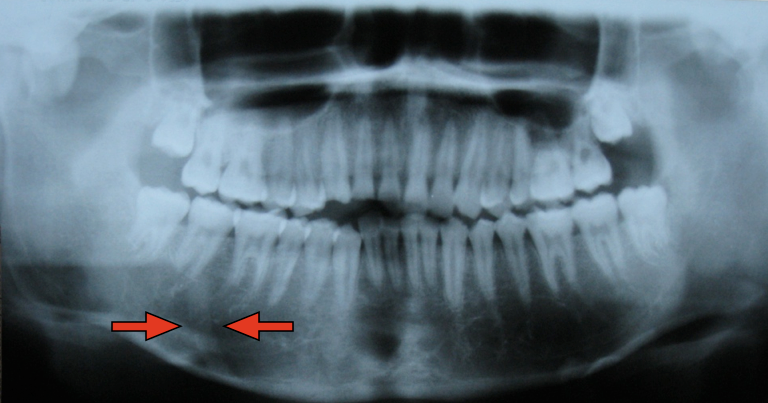
The next picture shows topographic view of the same case, the fracture fragment was overlapped from 44-47 area.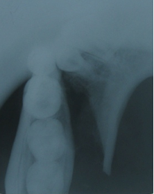
This last picture shows the fracture of right zygomatic arch. The arch was overlapped (in superior-inferior and medial-distal direction), thus it show more radiopaque and more widening than normal side.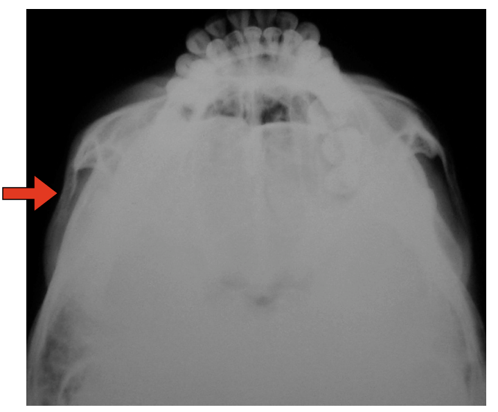
Relate posted
Showing posts with label facial fracture. Show all posts
Showing posts with label facial fracture. Show all posts
07 February 2009
x-ray and fracture line part 2
05 February 2009
x-ray and fracture line
The typical x-ray appearances of fracture included
- Radiolucent line
- Radiopaque line if the fragments overlie one another
- Discontinuity or deformity in the out line of the bone
Why can we see more radiopaque line in fracture look at this figure below?

Another example, in submentovertex view, in fracture zygomatic arch that the arch was collapsed and overlapped, show only slightly increase of radiopaque and widening of arch than contralateral arch.
Next time if I 'm not forget I will show the example of these film x-ray, however the most important in diagnosis the fracture of head and neck bone was not x-ray, but clinical.
Relate posted
29 January 2008
Diagnosis Image Facial Fracture
กระดูกบริเวณใบหน้านั้นมีความซับซ้อนค่อนข้างมาก การที่เราจำเป็นต้องตรวจผู้ป่วยที่ได้รับบาดเจ็บในบริเวณนี้นั้นจึงเป็นเรื่องไม่ง่ายนัก การรู้เพียงแค่ normal anatomy ของบริเวณนี้นั้นคงไม่พอเพียงเราจำเป็นต้องรู้ลึกไปถึง common facial fracture pattern อีกด้วย สำหรับบทความนี้ผู้เขียนได้บรรยายถึงการตรวจวินิจฉัย fracture ของกระดูกใบหน้าและ ขากรรไกร โดยเน้นไปที่การใช้ภาพรังสีในการตรวจ
สำหรับตัวผู้เขียนแล้วในสถาบันของเขาใช้ CT Scan ในการตรวจ fracture of Maxillofacial region เพื่อดูทั้ง hard +soft tissue (บ้านเราคงเอาอย่างเขายาก) ส่วน indication ในการใช้ plain film นั้นมีน้อยลงเรื่อยๆโดยมีการใช้ใน case ที่มี focal trauma เช่น Fracture nose แต่อย่างไรก็ตามผู้เขียนก็ยังเห็นความสำคัญของ plain film facial series ที่เราใช้เป็นพื้นฐานในการศึกษา normal anatomy และ Fracture pattern ในขั้นเริ่มต้นก่อนจะไปดู CT ใน plain ต่างๆ
Basic Facial series ประกอบด้วย 3-4 film คือ Water’s view(PA view with cephalad angulation) ,Caldwell’s view(PA view), Lateral view และ Submentovertex view ที่ได้ใช้ในบางโอกาส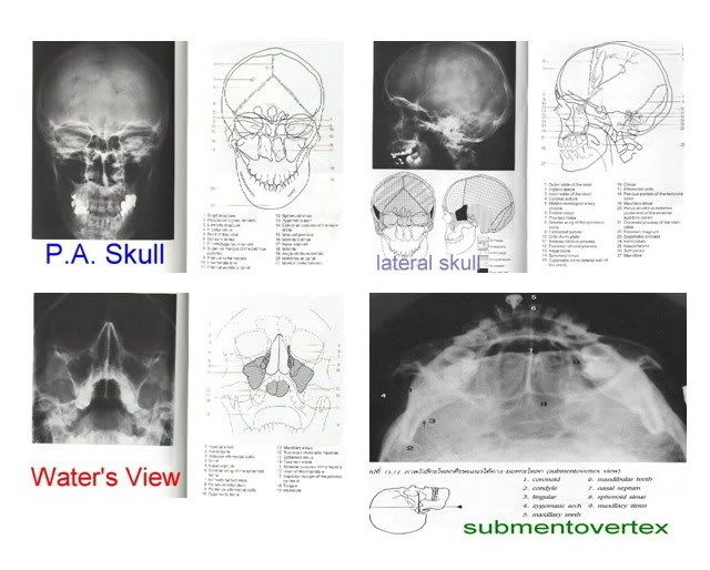
เอาละครับเมื่อถ่าย Film ต่างๆนี้มาแล้วก็มาถึงขั้นตอนการแปลผลหรืออ่าน film กันโดยมีกฎง่ายๆที่ผู้เขียนแนะนำไว้ดังนี้
- มองไปที่ Orbit ทั้งสองข้าง 60-70% ของ Facial fractures มักจะ involve orbit ด้วยแล้วแต่ว่าจะมากหรือน้อย จะมีที่ไม่มายุ่งกับ orbit ก็พวก fracture pure nasal bone ,fracture zygomatic arch, Lefort I fracture ฉนั้นอย่าลืมดูที่ orbit ให้ดีตั้งแต่ใน apex หรือ optic canal ออกมายัง orbital rim
- ข้อนี้บอกว่าเราต้องมีความรู้เกี่ยวกับ Facial fracture patterns เพื่อจะได้มีแนวในการสอดส่ายตา มองหาจุดที่หัก
- ใช้หลักการของการเปรียบเทียบ ซ้ายกับขวา เพราะมีหลักการที่เป็นความจริงเป็นภาษาอังกฤษที่ว่าใน Normal radiopacities are usually bilateral, while abnormal ones are usually unilateral
- ตรวจแนวเส้นรังสีของกระดูก Lines of Dolan จาก film Water’s view
Line of Dolan คือ เส้นในภาพรังสี ที่แสดง three anatomic contours ผ่าน facial important structure

หลังจากดูตามหลัก 4 ข้อไปแล้วเราก็เริ่มสังเกต Radiographic signs of Facial fractures กันต่อครับ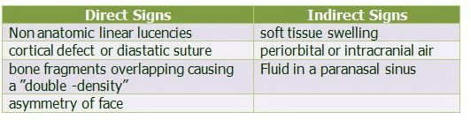
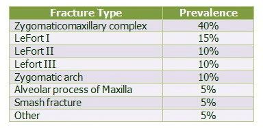
ตารางแสดง Most common patterns of midfacial fractures
(เป็น series ของ web แห่งนี้เท่านั้นนะครับ )
Article from http://www.geocities.com/omfsesan/facialfracture.htm
Relate links
Maxillary Fracture
Mandibular Fracture
Subscribe to:
Posts (Atom)
