04 November 2009

close reduction nasal bone

asch forcep


walsham forcep


Technique
Nasal pyramid fractures should be reduced first, followed by nasal septum reduction.
Explain the risks, benefits, and alternatives to the patient. Obtain a signed informed consent, if possible.
Deliver appropriate anesthesia. For details, see Anesthesia.
To reduce nasal pyramids, measure the distance from the alar rim to the depressed fragment externally. Mark position with thumb. Reduce depressed side of nose first.
Insert Boies or Salinger elevator into the nose under the depressed fragment. Apply steady outward pressure on the posterior aspect of the nasal bone. Control outward pressure with counterpressure exteriorly with the other thumb. Fragments may need to be molded into the proper position.

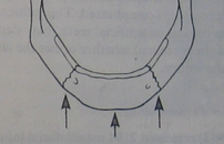
Nasal bone reduction.
If unable to reduce with elevators, use Walsham forceps to directly grasp the nasal bone. Insert one blade beneath the bone as the other blade is opposed on the outer skin surface. Manipulate the bone into position.
Check for septal reduction. If not adequately reduced, use Asch forceps to elevate the nasal pyramid while applying direct pressure to the displaced portion of the septum until it is moved back into the proper position.

Septal reduction with forceps.
Check for septal hematoma (drain if present).
Stabilize reduction with internal packing (eg, Vaseline gauze or 8-cm Merocel) and an external splint (eg, Thermaplast, Aquaplast). These external splints require intense heat for activation and molding, so the nasal dorsal skin should be protected with Steri-Strip bandage application prior to placement of the splint.
Remove packing in 5 days and remove nasal splint in 7 days. While the nasal packing is in place, the patient should be on an oral antibiotic with adequate Staphylococcus aureus coverage (eg, cephalexin) in order to prevent sinusitis and toxic shock syndrome.

27 September 2009

Ventilation of Lung alveoli


Ventilation ของ Alveoli ของปอดในแต่ละตำแหน่งนั้นไม่เท่ากัน โดย ventilation ในถุงลมปอดส่วนบนนั้นจะทำได้น้อยกว่าถุงลมปอดส่วนล่าง

เหตุใดจึงเป็นเช่นนั้น

เราลองนึกภาพของปอดสองข้างที่โดนแขวนอยู่ด้วยหลอดลม ดังนัั้นส่วนของ alveoli ของปอดล่างก็จะโดนน้ำหนักของเนื้อปอดส่วนบนทับลงมา ถุงลมของปอดส่วนบนจึงมีลมค้างอยู่มากกว่าถุงลมของปอดส่วนล่าง (ขณะสิ้นสุดการหายใจออก) นึกภาพให้ง่ายขึ้น ให้คิดถึงภาพของสปริงก็ได้ สปริงส่วนบนที่ยืดมากกว่าสปริงส่วนล่าง

ดังนั้นเราจึงสรุปได้ว่า ventilation ของ alveoli ของปอดล่างนั้นดีกว่าปอดบน เพราะว่าเมื่อหายใจเข้าถุงลมของปอดส่วนล่างจะขยายตัวได้มากกว่าเมื่อเทียบกับถุงลมของปอดส่วนบนที่ขยายตัวค่อนข้างมากอยู่แล้ว


19 August 2009

ยาลดความดันกลุ่ม calcium channel blocker

ยาลดความดันในกลุ่ม calcium channel blocker ทำงานโดยความรู้ที่ว่า ca2+ นั้นใช้ในการหดตัวตัวของกล้ามเนื้อดังนั้นเมื่อไป block มันแล้วก็จะทำให้ให้กล้ามเนื้อหดตัวได้น้อยลง จึงก่อให้เกิด vasodilatation ของ vessel และลดความแรงในการหดตัวของกล้ามเนื้อหัวใจ (negative ionotropic) ซึ่งต่างกับกลุ่ม beta channel blocker ที่ไปปิดกั้นที่สัญญาณไฟฟ้าที่ส่งมาที่หัวใจแต่ว่าความแรงในการบีบตัวของกล้ามเนื้อนั้นยังเท่าเดิม จากความรู้ที่กล่าวมาข้างต้นทำให้ผู้ป่วยที่ทานยา CCB มีผลข้างเคียงอย่างหนึ่งคือ tachycardia จากการที่ BP drop จากแรงบีบตัวของกล้ามเนื้อหัวใจที่ลดลง ทำให้ barorecepter ส่งสัญญาณกลับไปให้เพิ่มอัตราการเต้นของหัวใจ

จากความรู้เรื่อง action potential ใน cardiac muscle ทำให้รูได้ว่า mechanism อีกอย่างหนึ่งของ CCB คือจะยั้บยั้งการนำกระแสไฟฟ้าในหัวใจได้เช่นกัน ดังนั้นก็ทำให้มีโอกาสเกิด heart block ได้คล้ายๆกับ beta channel blocker โดยยากลุ่ม beta channel blocker นั้นจะ titrate ให้ได้ heart rate ที่ต้องการได้ง่ายกว่ากลุ่ม CCB

ยา CCB แบ่งเป็นอีก 3 กลุ่มคือ Dihydropyridine (-dipine),Phenylalkylamine(-pamil),Benzothiazepine(Diltiazem)
โดยยากลุ่ม dihydropyridine นั้นจะมีผลต่อ vessel มากกว่า ส่วนกลุ่ม phenylalkylamine จะมีผลต่อกล้ามเนื้อหัวใจมากกว่า ดังนั้นมักใช้เป็นยาพวก anti-angina ด้วย ส่วนกลุ่ม Benzothiazepine นั้นออกฤทธิ์ทั้งแต่ vessel และ cardiac

Hypertension and Stroke

cerebral blood flow mean arterial pressure autoregulation

จากกราฟจะเห็นว่ากระบวนการ autoregulation ของร่างกายอยู่ที่ mean arterial pressure ที่ 50-150 mmHg เมื่อไหร่ก็ตามถ้า MAP ลดลงต่ำกว่า 50 mmHg จะทำให้ Cerebral blod flow ที่ไปที่สมองไม่เพียงพอเกิดเป็น ischemic stroke ในทางกลับกันถ้า MAP > 150 ก็จะทำให้เกิด CBF ที่มากเกินไปเกิดเป็น hemorrhagic stroke ได้

autoregulation hypertension cerebral blood flow mean arterial pressure
มาดูอีกกราฟหนึ่งในผู้ป่วยที่มี hypertension จะพบว่า autoregulation interval นั้นเปลี่ยนไป โดย interval ใหม่ที่เกิดขึ้นจะแคบกว่าในคนปกติ ในคนไข้ hypertension นั้นความดันจะ swing ได้ค่อนข้างง่ายอยู่แล้ว เพราะฉะนั้นถ้าในกรณีทำหัตถการที่ทำให้คนไข้เครียดหรือเจ็บปวดก็จะทำให้ BP สูงเกิด hemorrhagic stroke กลับกันในกรณีในคนไข้ที่ทำหัตถการภายใต้การวางยาสลบ ถ้าให้ deep sedation มากเกินไปหรือยานำสลบที่ลดความดันมากเกินไปก็ทำให้เกิด ischemic stroke ตามได้เช่นกัน

เพื่อความเข้าใจมากขึ้นเข้าไปอ่านประกอบได้ที่นี้ http://medinfo2.psu.ac.th/anesth/education/trauma2.html

Intracranial Pressure and Cerebral Blood Flow

The brain is only able to withstand very short periods of ischaemia, unlike the kidney, liver or muscle

Maintenance of cerebral blood flow depends on a balance between the pressure within the skull, intracranial pressure (ICP) and the arterial pressure of the blood, mean arterial pressure (MAP).

Thus when blood pressure falls, physiological mechanisms attempt to maintain flow to prevent ischaemia. This process is autoregulation

If there is an increase in the volume of either the brain or blood the normal initial response is a reduction in CSF volume within the skull. CSF is forced out into the spinal sac

It is believed that a MAP that is greater than 60 mmHg is enough to sustain the organs of the average person.

CPP = MAP - ICP (normal 10 mmHg)
Normal cerebral perfusion pressure is 80 mmHg

but when reduced to less than 50 mmHg there is metabolic evidence of ischaemia and reduced electrical activity

In the head injured patient, CPP should not fall below 70 mmHg.

Drowsy and confused: (GCS 13-15)ICP=20 mmHg,Severe brain swelling (GCS <8) ICP=30 mmHg

The normal cerebral blood flow is 45-50ml 100g-1 min-1, ranging from 20ml 100g-1 min-1 in white matter to 70ml 100g-1 min-1 in grey matter. There are two essential facts to understand about cerebral blood flow. Firstly, in normal circumstances when the flow falls to less than 18-20ml 100g-1 min-1, physiological electrical function of the cell begins to fail

Normally autoregulation maintains a constant blood flow between MAP 50 mmHg and 150 mmHg

Carbon dioxide causes cerebral vasodilation. As the arterial tension of CO2 rises, CBF increases and when it is reduced vasoconstriction is induced.

Full article here http://www.nda.ox.ac.uk/wfsa/html/u08/u08_013.htm

14 August 2009

IV anesthetic agent

ยานำสลบที่ให้ทางหลอดเลือดดำมีด้วยกันหลายตัว เราพอจะมีขอพิจารณาคร่าวๆในการเลือกใช้ดังนี้


  • Thiopental ให้การนำสลบที่ค่อนข้าง smooth แต่อาจจะกระตุ้นให้เกิดการหลั่ง histamine ได้ thiopental มีฤทธิ์ vasodilatation แต่จะกระตุ้นให้ increase heart rate ทำให้ BP ไม่ drop มากนัก ถ้าให้ thiopental เป็นจำนวนมากจะทำให้ผู้ป่วยตื่นได้ช้า
  • Propofol นำสลบได้เร็ว และหมดฤทธิ์เร็ว มีผลให้ BP drop เนื่องจากมีฤทธิ์ vasodilatation แต่ไม่ compensate ด้วยการเพิ่ม Heart rate และยังทำให้เกิด apnea ได้ค่อนข้างสูง (30%) เวลาฉีด propofol ยังแสบอีกด้วยแนะนำให้ฉีดเข้าเส้นเลือดดำที่ค่อนข้างใหญ่จะดีกว่า
  • Ketamine มีฤทธิ์ในการนำสลบและ " แก้ปวด" ด้วย เหมาะในการนำสลบในคนไข้ที่อยู่ในภาวะ shock เพราะมีผลกระตุ้น ทั้ง HR และเพิ่ม muscle contraction ของหัวใจ ทำให้ BP สูง แต่การใช้ยานี้ทำให้เกิดลักษณะ dissociate anesthesia ได้
  • Etomidatel BP ค่อนข้าง stable ไม่ทำให้เกิดการหลั่ง histamine มีอาการระคายเคืองเวลาให้ยาเช่นเดียวกับ propofol


31 March 2009

action of lateral pterygoid muscle

sup head action when close mouth to couteract the force of retrodiscal pad, it create the smooth backward movement of menicus.

if the elastic of the retrodiscal pad is loose, the disc will protrude ant to the normal position.

PS. inf head of lat pterygoid will action when mouth openning

21 March 2009

diabetes ketoacidosis จะเกิดขึ้นได้เมื่อไหร่

Diabetes ketoacidosis ในคนไข้เบาหวานจะเกิดขึ้นเมื่อไหร่

ในคนไข้เบาหวานที่มีน้ำตาลในเลือดสูงมากๆ ถ้าไม่อยากให้น้ำตาลในเลือดสูง คิดง่ายๆเราก็ไม่ต้องให้คนไข้ได้น้ำตาลเลย คือไม่ให้ทานของหวานเลย ไม่ให้ fluid ที่มีน้ำตาลเป็นส่วนประกอบ ถ้าเราทำเช่นนั้น คนไข้เบาหวานจะเกิดภาวะ diabetes ketoacidosis ได้ ทำไมจึงเป็นเช่นนั้น ทั้งๆที่คนไข้ก็มีน้ำตาลในเลือดสูงอยู่แล้ว ที่เป็นเช่นนั้นก็เพราะว่า น้ำตาลในเลือดของคนไข้เบาหวานนั้นร่างกายไม่สามารถนำไปใช้เป็นพลังงานได้ แต่ถ้าเป็นน้ำตาลที่ metabolite ขึ้นมาใหม่นั้น cell จะสามารถนำไปใช้ได้

เพราะฉะนั้นถ้าเราไม่ให้น้ำตาลเลย ก็จะเกิด Diabetes ketoacidosis ได้ แล้วเราให้น้ำตาลได้เท่าไหร่ละถึงจะน้อยที่สุดที่จะไม่เกิดภาวะนี้ ต้องมาเข้าใจกระบวนการ physiology ของร่างกายก่อน

ร่างกายเราแบ่ง cell ในการใช้น้ำตาลได้ประมาณ 3 ประเภท
1. glucose dependent คือ สมอง และ RBC แปลว่า สมองจะไม่ใช้พลังงานในรูปอื่นนอกจาก glucose
2. glucose prefer คือพวก ตับ ไต
3. glucose independent คือไม่มีน้ำตาลก็ไม่เป็นไร เช่น พวก cell กล้ามเนื้อ, nerve cell ใช้พวก lactic acid ได้

เพราะฉะนั้น ถ้าสมองมีน้ำตาลไปเลี้ยงไม่พอ ร่างกายก็จะสลายไขมันเพื่อไปสร้างน้ำตาลมาใช้ ทำให้เกิดของเสียที่เรียกว่า ketone ออกมา นำไปสู่ diabetes ketoacidosis

แล้วสมองต้องใช้น้ำตาลต่อวันขัั้นต่ำเท่าไหร่ถึงจะเพียงพอละ คำตอบอยู่ที่ glucose 120 g แล้วมันเท่าไหร่ละ glucose 120 g นะ คิดง่ายๆ coke หนึ่งกระป๋องมีน้ำตาลอยู่ประมาณ 200 g เพราะฉะนั้นถ้าคนไข้กินโค้กได้วันละกระป๋องก็ไม่ต้องกลัวภาวะ diabetes ketoacidosis ล่ะ (แต่ถ้ากินโค้กด้วย กินข้าวด้วย กินผลไม้หวานๆด้วย น้ำตาลก็ไม่ลงเลยละคราวนี้ ต้องดูหลายๆอย่างประกอบ นี้แค่ยกตัวอย่างให้ดู)

เข้าไปอ่านต่อที่นี้แล้วจะเข้าใจมากขึ้น
http://www.absoluteastronomy.com/topics/Famine_response

19 March 2009

Regular insulin กับการควบคุมน้ำตาลในผู้ป่วยเบาหวานก่อนผ่าตัด

ในกรณีคนไข้ เบาหวานที่ต้องควบคุมระดับคนไข้เบาหวานก่อนผ่าตัด ส่วนใหญ่จะใช้ regular insulin ในการควบคุม

การให้ RI ให้ได้หลักๆสองทาง
1. IV ออกฤทธิ์ทันที peak ใน 30 นาที
2. subcutaneous ออกฤทธิ์ช้า peak ที่ 3 hr และมีผลต่อไปอีก 3 hr

โดย RI 1u จะสามารถลดน้ำตาลได้ 30-50 mg%
ถ้าคนไข้มี FBS > 200 จะ 1u จะลดได้ 50mg%
แต่ห้ามลด FBS > 100 mg% ใน 1 hr มิฉะนั้นจะเกิดภาวะสมองบวมจาก ภาวะที่ cell ได้น้ำตาลเข้าไปมากทำให้น้ำไหลตามน้ำตาลเข้าไปใน cell เกิด cell บวมขึ้น

ข้อควรรู้อีกอย่างคือ น้ำเกลือ 5%Dn/2 1 ขวด มีน้ำตาล 50 g ซึ่งต้องใช้ RI 10u ในการ metabolite ให้หมด ซึ่งก็คือ RI 1u metabolite glucose ได้ 5 g ความรู้นี้นำไปใช้ในการ titrated RI ผ่านทาง IV drip ได้

ข้อที่ควรนำมาพิจารณาเกี่ยวกับระดับน้ำตาลกับศัลยกรรม อีกอย่างหนึ่งคือ ระดับน้ำตาลหลังทำหัตถการเป็นเช่นไร เนื่องจากการทำ surgery จะทำให้เกิด trauma หลังทำ ทำให้ภาวะน้ำตาลสูงขึ้น แต่อีกทางหนึ่งถ้าคนไข้หลังทำหัตถการไม่สามารถกินได้ ระดับน้ำตาลในเลือดก็จะต่ำ ดังนั้นเราต้อง balance สองจุดนี้ให้ดี

สุดท้ายนี้ความรู้ด้านบนเป็นอัตรา RI ที่ลดน้ำตาลได้ในคนปกติ แต่ในคนเป็นเบาหวาน cell จะ response ต่อ RI ได้ไม่เหมือนคนปกติ ดังนั้นต้องทำการ titrated โดยปรับ dose ไปเรื่อยๆแล้วเจาะ Dtx เป็นระยะเผื่อประเมินว่าในคนไข้คนนี้ reponse ต่อ RI อย่างไร

11 March 2009

staging of osteoradio necrosis (ORN)

osteoradionecrosis เราสามารถแบ่งออกเป็นทั้งหมด 3 stage โดย stage III ตามตัวหนังสือที่เขียนไว้ก็คือ ลักษณะของ ORN ร่วมกับมี fistula opening หรือมี pathologic fracture ของตัวกระดูกร่วมด้วย ซึ่งถ้าเราแบ่ง stage ของ ORN ตามลักษณะของมันนั้น เราจะยังเข้าไม่ถึงหัวใจของการแบ่ง ORN

อธิบายอีกทางก็คือ สมมุติเราพบ ORN ที่มี fistula openning แต่เราส่งไปทำ HBO แล้วอาการผู้ป่วย response ดีขึ้นจนหายดีโดยเราไม่ต้องทำ intervention ใดๆเลย เรายังจะให้เป็น ORN stage III หรือไม่

ดังนั้น จริงๆแล้วการแบ่ง stage ORN อย่างง่ายๆเลยก็คือ ถ้า ORN ที่ response ต่อ HBO 3o drive แล้วสามารถรักษาหายได้โดยไม่ต้องทำ local debridement ใด เราถือว่าเป็น stage I แต่ถ้าได้ HBO 30 drive อาการไม่ดีขึ้น ต้องทำ local debridement แล้วรักษาหายได้ เราถือว่าเป็น ORN stage II และสุดท้ายถ้ารักษาด้วยการ local debridement ไม่หาย ต้องใช้การรักษาที่ aggressive มากขึ้นไปอีก เราก็จะถือว่ามันเป็น ORN stage III

21 February 2009

oculocardiac reflex

In maxillofacial surgery, the oculocardic reflex can be occur when we explore orbit or any operation that have to associated with orbit.

-----------------------

The oculocardiac reflex, also known as Aschner phenomenon, Aschner reflex, or Aschner-Dagnini reflex, is a decrease in pulse rate associated with traction applied to extraocular muscles and/or compression of the eyeball. The reflex is mediated by nerve connections between the trigeminal cranial nerve and the vagus nerve of the parasympathetic nervous system. The afferent tracts derive mainly from the ophthalmic division of the trigeminal nerve, although tracts from the maxillary and mandibular division have also been documented[1]. These afferents synapse with the visceral motor nucleus of the vagus nerve, located in the reticular formation of the brain stem. The efferent portion is carried by the vagus nerve from the cardiovascular center of the medulla to the heart, of which increased stimulation leads to decreased output of the sinoatrial node[2]. This reflex is especially sensitive in neonates and children, and must be monitored, usually by an anaesthesiologist, during paediatric ophthalmological surgery, particularly during strabismus correction surgery[3]. However, this reflex may also occur with adults. Bradycardia, junctional rhythm, asystole, and very rarely death [4], can be induced through this reflex.

Treatment/prophylaxis

Removal of the inciting stimulus is immediately indicated, and is essential for successful termination of this reflex. The surgeon, or practitioner, working on the eye should be asked to cease their activity and release the applied pressure or traction on the eyeball. This often results in the restoration of normal sinus rhythm of the heart. If not, the use of an anti-muscarinic acetylcholine (ACh) antagonist, such as atropine or glycopyrolate, will likely successfully treat the patient and permit continuation of the surgical procedure. In extreme cases, such as the development of asystole, aggressive cardiopulmonary resuscitation may be required.

20 February 2009

Lefort I osteotomy video clip

I found this lefort I osteotomy clip in youtube.

19 February 2009

fracture mandible and airway obstruction

Fracture mandible that will increase the risk of airway obstruction mostly appears in 2 form.

  1. Fracture bilateral symphysis of mandible
  2. Fracture bilateral condyle of mandible

fracture bilateral symphysis of mandible

First, the symphysis acted like free segment (coz fracture bilat.), suprahyoid muscle (ex. anterior belly of digastric) will pull this segment back, The oral cavity appeared smalller, tongue have no space to occupy. This sequence make patient difficult to breath.

suprahyoid muscle

Latter, the fracture of bilateral condyle of mandible, in this type when patient lie down the mandible will retrude. In the same sequence, tongue have no space to occupy and patient is hard to breath.

Thus the patients in these two type (maybe) can't lie in supine position, the best position is lie on one side or semi supine position to make sure the fracture fragment will not retrude posterior.

18 February 2009

ANATOMIC CONSIDERATIONS IN NASAL BONE FRACTURE PART II: Blood supply

The rich blood supply to the nasal region predisposes individuals to epistaxis in nasal trauma. Epistaxis can be categorized as anterior or posterior, depending on the source of bleeding. The most common location is anterior epistaxis, originating from Kiesselbach’s plexus in the anteroinferior septum, which receives its blood supply from both internal and external carotid arteries. Posterior epistaxis typically arises from branches of both the sphenopalatine and anterior ethmoidal arteries. It is less common than anterior bleeding but a more frequent cause of severe hemorrhage. Nasal hemorrhage can usually be controlled by direct manual pressure in anterior bleeds. Hemorrhage from posterior sources usually resolves spontaneously, particularly if the patient’s blood pressure is controlled. However, in persistent cases, posterior packing or Foley balloon catheterization of the nasal passage may be necessary. In the most severe cases, usually secondary to maxillary arterial sources, interventional embolization may be required. Both the ophthalmic and maxillary divisions of the trigeminal nerve supply sensation to the nose. The infratrochlear nerve provides sensation to the skin of the upper nasal dorsum and sidewalls, and the anterior ethmoidal supplies the lower dorsum and tip.
nasal blood supply

17 February 2009

ANATOMIC CONSIDERATIONS IN NASAL BONE FRACTURE PART I: Bony part

The skeletal component of the nose includes the frontal process of the maxilla, the nasal process of the frontal bone, the ethmoid, the vomer, and the nasal bones. Fractures of the nasal bones occur more commonly distally, where they are broader and thinner. Significant force is required to fracture the more proximal nasal bones, in which case injury extension into the frontal process of the maxilla and frontal bone may be present.
The cartilaginous structures include the two lower lateral cartilages, two upper lateral cartilages, and the central septal cartilage. Paired upper lateral cartilages have solid attachments to the caudal aspect of the nasal bones and help maintain the midline location of the quadrangular cartilage. The lower lateral cartilages provide little midline
structural support but are responsible for much of the aesthetics and contour of the nasal tip. The fact that cartilage surrounds the bony matrix both dorsally and laterally to provide a soft and malleable framework allows some dissipation of force without incurring a permanent deformity. In general, significant force is required for a cartilaginous
injury.
nasal bone
nasal septum

12 February 2009

temporal branch of facial nerve injury (danger zone)

The temporal branch of the facial nerve is at greatest risk for injury where it crosses the zygomatic arch. Injury to the temporal branch of the facial nerve leads to an inability to elevate the eyebrows and brow ptosis and paralysis. Asymmetric appearance of the forehead can also occur because there will be a loss of the lines and wrinkles on the affected side. One easy method to delineate the danger area of the temporal nerve is to draw a line from the earlobe to the lateral edge of the eyebrow, and the tragus to just above and behind the highest forehead crease(or 2 cm above supraorbital rim). In the area between these lines , the nerve is superficial, and it is critical to undermine just below the dermis in the superficial fat above the fascia.


Relate posted

07 February 2009

x-ray and fracture line part 2

This post will show the radiographic sign that I tell you in x-ray and fracture line posted

The figure below shows the oblique fracture that the fracture fragment was oblique and overlapped in bucco-lingual direction, thus, show the more radiopaque line (red arrow)

oblique fracture mandible

The next picture shows topographic view of the same case, the fracture fragment was overlapped from 44-47 area.

oblique fracture mandible

This last picture shows the fracture of right zygomatic arch. The arch was overlapped (in superior-inferior and medial-distal direction), thus it show more radiopaque and more widening than normal side.
fracture zygoma

Relate posted

05 February 2009

x-ray and fracture line

The typical x-ray appearances of fracture included

  1. Radiolucent line
  2. Radiopaque line if the fragments overlie one another
  3. Discontinuity or deformity in the out line of the bone
The second is the most miss point when we interpreted film x-ray.
Why can we see more radiopaque line in fracture look at this figure below?

xrayfracture

For example, the oblique fracture of mandible ,we will didn't notice in x-ray because it sometimes presented only slightly not notably more radiopaque line. (The fracture fragment was overlapped)

Another example, in submentovertex view, in fracture zygomatic arch that the arch was collapsed and overlapped, show only slightly increase of radiopaque and widening of arch than contralateral arch.

Next time if I 'm not forget I will show the example of these film x-ray, however the most important in diagnosis the fracture of head and neck bone was not x-ray, but clinical.


Relate posted

04 February 2009

What is osteoradionecrosis (ORN) ?

Osteoradionecrosis easy definition is

1. Patient who previously recieve radiotherapy > 5000 rad
2. Bone exposed with non healing > 3 month

Stage of Osteoradionecrosis -- ORN

Osteoradionecrosis (ORN) can divided into 3 stage

Stage 1 is superficial osteoradionecrosis that can resolve with irrigation and hyperbaric oxygen (HBO)
In stage 1 osteoradionecrosis no bone surgical removal is required.

Stage 2 is stage 1 that progressive and can't healing with treatment method in stage 1

Stage 3 is ORN that have orocutaneous fistula, Pathologic fracture or resorption of inferior border of mandible.

We have to known each stage of ORN for effective treatment protocol that I show below
osteoradionecrosis treatment protocol

For full description of 3 stage of osteoradionecrosis read below.

-----------------------------------------------

ORN Staging System

Stage I
All patients who meet the definition of osteoradionecrosis except those with cutaneous
fistulae, pathologic fracture, or radiographic evidence of bone resorption. (These three
subsets of patients are classified as Stage III.) Stage I patients receive 30 HBOT
treatments. Wounds are maintained with irrigation only or with saline rinses. No bone is
surgically removed. If, after 30 treatments, the wound shows improvement, as evidenced
by decrease in amount of exposed bone; less resporption or spontaneous sequestration of
exposed bone; or decreased softening of exposed bone; the patient completes another 10
treatments. The total 40 HBOT treatments are meant to achieve full mucosal cover. If
there is no clinical improvement after 30 HBOT treatments or complete resolution, such
as extended or continued exposure of bone, absence of mucosal proliferation, or presence
of inflammation, the patient is advanced further to Stage II. (See Technical Treatment
Protocol for suggestions on pressure levels and duration of individual treatments.)

Stage II
Surgeons attempt a local surgical debridement to identify those patients with only
superficial or cortical bone involvement, and who can be resolved without jaw resection.
A transoral alveolar sequestrectomy is performed with fine saline-cooled, air-driven
saws. Minimal periosteal manipulation is done for labial access to the mandible. The
lingual periosteum is allowed to remain completely attached until the specimen is
delivered, and then only that which is attached to the specimen is reflected from bone.
The resultant mucoperiosteal flaps are closed in three layers over a base of bleeding bone.

If healing progresses without complication, HBOT is continued with 10 additional
sessions. If the wound dehisces, leaving exposed non-healing bone, the patient isadvanced to Stage III. In those patients who initially present with orocutaneous fistulae,
with pathologic fracture, or with radiographic osteolysis to the inferior border, the initial
30 HBOT treatments are given, and the patient enters Stage III directly.

Stage III
After 30 HBOT treatments, the patient undergoes a transoral partial jaw resection, the
margins of which are determined at the time of surgery by the presence of bleeding bone. The segments of the mandible are stabilized either with extraskeletal pin fixation or with maxillo mandibular fixation. If there was an orocutaneous fistula, or large soft tissue loss, primary closure or soft-tissue reconstruction is accomplished during this surgery. HBOT is continued for another 10 treatments, and the patient is advanced to Stage III-R.

Stage III-R
These patients present the most difficult cases. Early reconstruction and rehabilitation
produces the best results. Ten weeks after resection, the soft tissues are healing, and the potential graft bed is free from contamination and infection. Bony reconstruction is
undertaken from a strictly transutaneous approach without oral flora contamination.
Ten post-reconstructive HBOT treatments are given, and the jaw fixation is maintained
for eight weeks. If no further surgery is required, the patient begins appointments with a
maxillofacial prosthodontist for full prosthetic rehabilitation one month after release of
his fixation. If additional surgery is required, such as a tongue release, vestibuloplasty, or excision of redundant tissue, surgeons schedule procedures one month after fixation is released. After surgery, the patient is referred to a maxillofacial prosthodontist.

02 February 2009

Tear lacrimal canaliculi injury

Lacrimal canaliculi injury can be occur in head and neck injury and have to specially suspected in patient that have laceration wound at eyelid involve lid margin.

See the picture of lacrimal duct system, the vertical part of canaliculi about 2 mm and horizontal part about 8 mm, thus the laceration wound in range about 5-10 mm from medial palpebral fissure have highly incident of lacrimal canaliculi injury. (the injury that medial to the punctum of eye.)
lacrimal canaliculi

Lacrimal canaliculi have both at upper and lower lid margin, the openning is through the upper and lower punctum. The percent of tear drainage are 75% from lower canaliculi and remaining for upper canaliculi. The main is at lower lacrimal canaliculi. If it have injury to this canaliculi and we didn't repair it the tear will can't drain and epiphora will occur.


punctum lacrimal canaliculi


Next post I will show method to repair canaliculi.
See ya.

01 February 2009

CA node metastasis or not

How to know this is CA metastasis node or not?

  1. Found primary CA
  2. Fix node (You have to try move node in every direction to confirm it fix or not)
  3. Node diameter > 1 cm
  4. Chain node (In viral infection also have chain node, but viral infection will have prodormal sign)
  5. Node was no tender (In general, infection node will have tenderness, anyway, chronic infection will also absence of tenderness.)
This is not the rules and the CA metastasis node haven't to have all this criteria.
It just the consideration point to determine that is it CA metastasis node or not.

31 January 2009

Melanocyte melanin pigmented lesion

Melanin lesion or hyper-pigmented occurred on the skin or mucosa when melanocyte had produce more melanin pigment than normal.

Why?

3 factor that involve in increase melanin pigment

  1. Genetic
  2. UV
  3. Hormone
The first one "Genetic"
Look at the picture!
Roy keane and Viera have differed skin color, the difference is at the ability of the melanocyte to produce melanin not the number of melanocyte(This 2 people have same number of melanocyte), which this ability is controled by Genetic.

melanin


Second "UV"
UV will convert tyrosine to tyrosinase and transform to Dopa and finally product is melanin pigment. This is the reason that why UV make us hyper-pigmented. But if you are the white one, maybe you will not blacken but your skin will turn red instead. Because the color of skin is not only control by UV but the genetic is also influence.

tyrosine melanin


The last "Hormone"
Let me told about the some basic of hormone that involve in produce the melanin pigment first. Melanin stimulating hormone (MSH) is control the production of melanin pigment, MSH is secreated by anterior pituitary gland, same as adrenocorticotropic hormone(ACTH) and Tyroid stimulating hormone(TSH).

The research discover that MSH shares the same precursor molecule as ACTH, so if your body generate more ACTH, your MSH also elevated.
For example, the Addison disease (chronic adrenal insufficiency) patient who can't secrete cortisol hormone. Thus, Anterior pituitary gland(negative feedback from low level of cortisol hormone) will produce more ACTH to stimulate adrenal gland to secrete cortisol. The side effect is also produced more MSH. It is why Addison disease patient have hyper-pigmented lesion at skin.

ACTH MSH melanin


Relate posted

23 January 2009

White and red oral lesion

Basic principle to know before deal with red and white lesion in oral mucosa
is how come it represent in white or red.

Back to the normal anatomy of oral mucosa as this pic.
white oral lesion

White zone compose with epithelium and lamina propria
and red zone is at submucosal area that has vascular.

Back to the same question how come it white, thinking to the white zone, what will increase white zone and make white zone more notable.

  1. Thickening layer of keratin
  2. Hyperplasia of epithelium
  3. Intracellular edema
  4. Decrease vascular at submucosal layer
  5. and other stuff cover the surface such as necrotic tissue, candida
Thus, we also can group white lesion into scraped off and cannot scraped group.
1-4 are cannot wiped off group and 5 is wiped off group.
white oral lesion

Back to the red lesion, how come it red, same logic, thought back to the red zone.
What does make it prominent?

1. Thinning or atrophy of white zone (epithelium, lamina propria) so red zone will show more notable.
2. Increase in vascular in red zone such as inflammation, bleeding, increase hemoglobin concentration (polycytemia vera)

Back to anatomy and physiology.
Good luck.

Related post

20 January 2009

Hemostatic agent in dental use

Hemostatic agent ที่ใช้บ่อยทางทันตกรรมและ Maxillofacial

1.Surgicel
Surgicel เป็น acidic material ซึ่งก็คือ oxidized cellulose มันจะทำปฎิกริยากับเลือดทำให้เกิด clot สีน้ำตาลแดง ซึ่งเป็นลักษณะของ pseudoclot สามารถทำให้เลือดหยุดไหลได้ surgicel เป็น absorable material สามารถสลายไปเองได้

2. Bone wax
Bone wax สามารถ stop bleed ได้ด้วย tamponade effect (การหยุดเลือดด้วย outer force ทำให้เกิดการ constrict ของ vessel) มีประสิทธิภาพที่ดี ใช้ง่าย แต่มีข้อเสียค่อนข้างมากเนื่องจากจะไปเคลือบผิว bone ทำให้

  • bone ไม่สามารถมี osteogenesis เนื่องจาก bone wax ไปเคลือบผิว bone ไว้ ทำหน้าที่เป็น physical barrier ทำให้ osteoblast ไม่สามารถวิ่งเข้ามาได้ หรือว่าในกรณี bone fracture ก็จะไม่สามารถกลับมา union กันได้เพราะถูก bone wax เคลือบอยู่
  • bone wax ยังเพิ่ม rate of infection เนื่องจากมันเป็น foreign body
3. Gelfoam
Gelfoam เป็น absorable gelatin sponge มีผลทาง physical โดยจะเป็นโครงให้ fibrin มาเกาะ

4. Fibrin glue
fibrin glue เป็นสารสังเคราะห์ประกอบด้วย fibrinogen และ thrombin เมื่อรวมกันแล้วทำให้เกิด fibrin และ develope ไปเป็น fibrin clot ต่อไป

fibrin glue


5. Tranexamic acid

tranexamic acid เป็นสารที่ขัดขวางไม่ให้ plasminogen เปลี่ยนเป็น plasmin (plasmin จะทำให้ fibrin clot สลายไป) โดย transamin จะให้ 250mg 1*3
Tranexamic

19 January 2009

Bacteria in sinusitis

  1. Strep. pneumonia Gram positive cocci
  2. H. influenza Gram negative bacilli
  3. Moraxella Catarrhalis Gram negative cocci
เชื้อทั้งสามชนิดสามารถฆ่าได้ด้วย Amoxycillin ดังนั้น amoxycillin จึงเป็น drug of choice ของการ treat sinusitis แต่ถ้าผู้ป่วยได้รับการ treat มาแล้วหรือว่าทาน amoxy มาเป็นประจำอาจทำให้เกิดพัฒนาของเชื้อที่สามารถผลิต beta lactamase ได้ จึงอาจต้องเปลี่ยนยาเป็น Augmentin

18 January 2009

Biopsy

หลักการในการเลือกตำแหน่งที่จะทำ biopsy

1. represent lesion
เลือกตำแหน่งที่สามารถ represent lesion ได้
2. think one or two step forward เสมอ
ต้องคิดก่อนว่ารอยโรคนี้น่าจะต้องรักษายังไง partial mandibulectomy with bone graft หรือ enucleation เนื่องจากการเปิด flap เพื่อทำ biopsy มีความหมายถ้าต้องทำ bone graft ต้องเลือกการเปิด flap ที่ต้องไม่ compromise graft ในภายหลัง (เช่น การไม่ทำ vertical incision ในการเปิด flap)