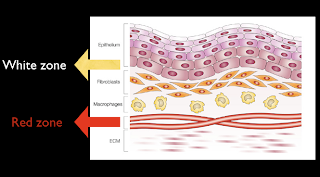
Basic principle to know before deal with red and white lesion in oral mucosa
is how come it represent in white or red.
Back to the normal anatomy of oral mucosa as this pic.
White zone compose with epithelium and lamina propria
and red zone is at submucosal area that has vascular.
Back to the same question how come it white, thinking to the white zone, what will increase white zone and make white zone more notable.
- Thickening layer of keratin
- Hyperplasia of epithelium
- Intracellular edema
- Decrease vascular at submucosal layer
- and other stuff cover the surface such as necrotic tissue, candida
1-4 are cannot wiped off group and 5 is wiped off group.

Back to the red lesion, how come it red, same logic, thought back to the red zone.
What does make it prominent?
1. Thinning or atrophy of white zone (epithelium, lamina propria) so red zone will show more notable.
2. Increase in vascular in red zone such as inflammation, bleeding, increase hemoglobin concentration (polycytemia vera)
Back to anatomy and physiology.
Good luck.
Related post
No comments:
Post a Comment