31 January 2009

Melanocyte melanin pigmented lesion

Melanin lesion or hyper-pigmented occurred on the skin or mucosa when melanocyte had produce more melanin pigment than normal.

Why?

3 factor that involve in increase melanin pigment

  1. Genetic
  2. UV
  3. Hormone
The first one "Genetic"
Look at the picture!
Roy keane and Viera have differed skin color, the difference is at the ability of the melanocyte to produce melanin not the number of melanocyte(This 2 people have same number of melanocyte), which this ability is controled by Genetic.

melanin


Second "UV"
UV will convert tyrosine to tyrosinase and transform to Dopa and finally product is melanin pigment. This is the reason that why UV make us hyper-pigmented. But if you are the white one, maybe you will not blacken but your skin will turn red instead. Because the color of skin is not only control by UV but the genetic is also influence.

tyrosine melanin


The last "Hormone"
Let me told about the some basic of hormone that involve in produce the melanin pigment first. Melanin stimulating hormone (MSH) is control the production of melanin pigment, MSH is secreated by anterior pituitary gland, same as adrenocorticotropic hormone(ACTH) and Tyroid stimulating hormone(TSH).

The research discover that MSH shares the same precursor molecule as ACTH, so if your body generate more ACTH, your MSH also elevated.
For example, the Addison disease (chronic adrenal insufficiency) patient who can't secrete cortisol hormone. Thus, Anterior pituitary gland(negative feedback from low level of cortisol hormone) will produce more ACTH to stimulate adrenal gland to secrete cortisol. The side effect is also produced more MSH. It is why Addison disease patient have hyper-pigmented lesion at skin.

ACTH MSH melanin


Relate posted

23 January 2009

White and red oral lesion

Basic principle to know before deal with red and white lesion in oral mucosa
is how come it represent in white or red.

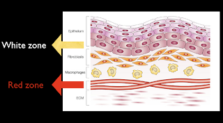
Back to the normal anatomy of oral mucosa as this pic.
white oral lesion

White zone compose with epithelium and lamina propria
and red zone is at submucosal area that has vascular.

Back to the same question how come it white, thinking to the white zone, what will increase white zone and make white zone more notable.

  1. Thickening layer of keratin
  2. Hyperplasia of epithelium
  3. Intracellular edema
  4. Decrease vascular at submucosal layer
  5. and other stuff cover the surface such as necrotic tissue, candida
Thus, we also can group white lesion into scraped off and cannot scraped group.
1-4 are cannot wiped off group and 5 is wiped off group.
white oral lesion

Back to the red lesion, how come it red, same logic, thought back to the red zone.
What does make it prominent?

1. Thinning or atrophy of white zone (epithelium, lamina propria) so red zone will show more notable.
2. Increase in vascular in red zone such as inflammation, bleeding, increase hemoglobin concentration (polycytemia vera)

Back to anatomy and physiology.
Good luck.

Related post

20 January 2009

Hemostatic agent in dental use

Hemostatic agent ที่ใช้บ่อยทางทันตกรรมและ Maxillofacial

1.Surgicel
Surgicel เป็น acidic material ซึ่งก็คือ oxidized cellulose มันจะทำปฎิกริยากับเลือดทำให้เกิด clot สีน้ำตาลแดง ซึ่งเป็นลักษณะของ pseudoclot สามารถทำให้เลือดหยุดไหลได้ surgicel เป็น absorable material สามารถสลายไปเองได้

2. Bone wax
Bone wax สามารถ stop bleed ได้ด้วย tamponade effect (การหยุดเลือดด้วย outer force ทำให้เกิดการ constrict ของ vessel) มีประสิทธิภาพที่ดี ใช้ง่าย แต่มีข้อเสียค่อนข้างมากเนื่องจากจะไปเคลือบผิว bone ทำให้

  • bone ไม่สามารถมี osteogenesis เนื่องจาก bone wax ไปเคลือบผิว bone ไว้ ทำหน้าที่เป็น physical barrier ทำให้ osteoblast ไม่สามารถวิ่งเข้ามาได้ หรือว่าในกรณี bone fracture ก็จะไม่สามารถกลับมา union กันได้เพราะถูก bone wax เคลือบอยู่
  • bone wax ยังเพิ่ม rate of infection เนื่องจากมันเป็น foreign body
3. Gelfoam
Gelfoam เป็น absorable gelatin sponge มีผลทาง physical โดยจะเป็นโครงให้ fibrin มาเกาะ

4. Fibrin glue
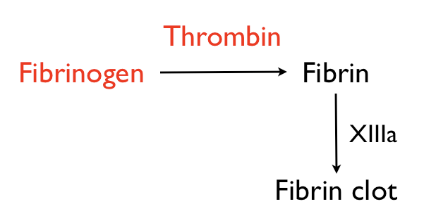
fibrin glue เป็นสารสังเคราะห์ประกอบด้วย fibrinogen และ thrombin เมื่อรวมกันแล้วทำให้เกิด fibrin และ develope ไปเป็น fibrin clot ต่อไป

fibrin glue


5. Tranexamic acid

tranexamic acid เป็นสารที่ขัดขวางไม่ให้ plasminogen เปลี่ยนเป็น plasmin (plasmin จะทำให้ fibrin clot สลายไป) โดย transamin จะให้ 250mg 1*3
Tranexamic

19 January 2009

Bacteria in sinusitis

  1. Strep. pneumonia Gram positive cocci
  2. H. influenza Gram negative bacilli
  3. Moraxella Catarrhalis Gram negative cocci
เชื้อทั้งสามชนิดสามารถฆ่าได้ด้วย Amoxycillin ดังนั้น amoxycillin จึงเป็น drug of choice ของการ treat sinusitis แต่ถ้าผู้ป่วยได้รับการ treat มาแล้วหรือว่าทาน amoxy มาเป็นประจำอาจทำให้เกิดพัฒนาของเชื้อที่สามารถผลิต beta lactamase ได้ จึงอาจต้องเปลี่ยนยาเป็น Augmentin

18 January 2009

Biopsy

หลักการในการเลือกตำแหน่งที่จะทำ biopsy

1. represent lesion
เลือกตำแหน่งที่สามารถ represent lesion ได้
2. think one or two step forward เสมอ
ต้องคิดก่อนว่ารอยโรคนี้น่าจะต้องรักษายังไง partial mandibulectomy with bone graft หรือ enucleation เนื่องจากการเปิด flap เพื่อทำ biopsy มีความหมายถ้าต้องทำ bone graft ต้องเลือกการเปิด flap ที่ต้องไม่ compromise graft ในภายหลัง (เช่น การไม่ทำ vertical incision ในการเปิด flap)