- Fracture Zygoma (Tripod Fracture) น่าจะเป็น fracture ที่เกิดขึ้นมากที่สุดในบรรดา fracture of facial bone pattern ของ fracture มักจะเกิดจาก direct blow to body of zygoma จากการตรวจจะพบเห็นลักษณะของ contour abnormalities,มีอาการของ extraocular muscle entrapped (คือมี limitation of eye movement ในบางทิศทาง) และ force ที่มาจากทาง lateral wall of orbit อาจส่งผลไปถึง orbital apex ,optic canal เกิด diminish vision ได้ ,นอกจากนั้นการ displace ไปของ zygoma อาจไปกีดกัน motion of mandible
ส่วนในภาพรังสี x-ray โดยเฉพาะใน water’s view จะเห็น discontinuity lines of Dolan และแน่นอนครับ ผู้เขียนแนะนำว่า CT จะช่วยการ diagnosis ได้ดีมาก
- Fracture Zygomatic arch pattern of fracture นั้นเกิดจากมีแรงมากระทำทางด้านข้างของ ใบหน้า ลักษณะของใบหน้านั้นจะพบว่ามี flatness of lateral cheek area and inability to open mouth อันเนื่องมาจากการที่ zygomatic arch fragment ลงมาขัดขวางการเคลื่อนที่ของ coronoid process และ temporalis muscle ส่วนภาพรังสีที่ใช้ในการ diagnosis ได้ดีคือ view submentovertex

- Fracture of Alveolar process of Maxilla เกิดขึ้นกับ small piece of maxilla ร่วมกับมี several fracture teeth สำหรับข้อ comment เกี่ยวกับ x-ray นั้นมีอยู่ว่าในกรณีที่สงสัย film chest x-ray จะช่วยในการตรวจสอบว่ามีการ aspirate foreign body หรือ tooth fragment ลงไปใน airway หรือไม่
- Orbital floor fracture , or “Blowout” fracture mechanism ของ fracture ก็คือว่า มีแรงหรือวัตถุมากระแทกรอบๆบริเวณดวงตา แล้วส่งแรงผ่าน soft tissue กระจายไปรอบๆดวงตาและผ่านมายัง thin floor of orbit เกิดการ fracture floor orbit และมี dropdown of orbital content to maxillary sinus
clinical sign ที่พบได้ก็คือ enophthalmos and diaplopia ส่วนใน film Water’s view จะพบเงาของ soft tissue mass บริเวณ superior margin of maxillary sinus หรืออาจจะเห็น trapdoor fragment of bone protruding down to sinus
- Fracture Nose โดยปกติแล้วเราสามารถเห็น fracture นี้ได้จาก standard lateral skull film แต่ถ้าเราใช้ technique low kVp ใน x-ray ก็จะสามารถเห็น บริเวณ fracture นั้นและ soft tissue ได้ดีขึ้น แต่ก็ต้องระวังการดู film ผิดพลาดคือเห็น normal suture line หรือ linear channel for Nasociliary nerve เป็น fracture line ได้เหมือนกันนะครับ เราควรจะดูใน lateral film ที่มีแนวรังสีตั้งฉากกับ nose จะทำให้เห็น fracture line ที่มักจะเป็นแนวตั้งฉากกับ nasal bridge ได้ชัดเจนกว่า

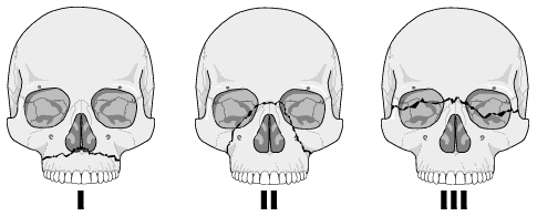
- LeFort fracture หรือเป็น fracture ที่ complex bilateral fractures associated with large unstable fragment and invariably involve pterygiod plates โดยแนวของ fracture นั้นแบ่งเป็น 3 ระดับคือ
- LeFort I มีแนว Fracture ตาม transmaxillary plane
- LeFort II มีแนว fracture ตาม subzygomatic หรือ pyramidal plane ที่มี apex บริเวณ nasal bridge
- LeFort III มีแนว fracture ตาม craniofacial plane
- Smash Fracture มีการคิดคำนี้ขึ้นมาเพื่อใช้เรียก severe comminution of the face with underlying skull injuries และยังรวมไปถึง fracture facial bone ที่ไม่สามารถ classified ไว้ในกลุ่มที่ผ่านๆมาได้ โดยรวมไปถึง Fracture frontal ,naso-ethmoidal ,naso-frontal ซึ่ง Fracture ในกลุ่มนี้ควรใช้ CT ในการตรวจจะได้รายละเอียดที่ดีกว่า plain film ทั่วไป
6.1 LeFort I หรือ tranmaxillary fracture มีแนวหัก ไปตาม maxillary floor & orbital floor โดย involve medial & lateral walls of maxillary sinuses และทางด้านหลัง involve pterygoid plate of sphenoid bone จะพบลักษณะ floating maxilla
6.2 Lefort II หรือ เรียกตามแนวทางของ fracture line ว่า Pyramidal fracture ลักษณะของ fracture นี้หลังจากมีแรงในแนวตรงเข้าด้านหน้ามากระทำแล้วจะมี mechanism ที่ downward below to nasal area
6.3 LeFort III เป็น most severe ของ LeFort fracture ลักษณะจะเป็น Large unstable(floating) fragment ทั่วไปทั้งใบหน้า หรือเรียกตามอาการว่า Craniofacial disassociation
Relate links
Diagnosis Image Facial Fracture
Mandibular fracture

No comments:
Post a Comment